Completed Research Project Listings:
The Computer Vision and Image Analysis group at Cornell University is involved in various research projects with the primary goal of the development of medical computer aided diagnosis systems. Other ongoing projects also are involved in the creation of programs and systems that can be used in a research setting. The following is a listing of ongoing graduate research projects and completed projects. All projects in VIA group are under the supervision of A.P. Reeves: Computer Vision and Medical Image Analysis Overview
VIA Research Projects:
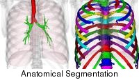 |
Automated Chest Health Analysis Automated chest health analysis involves the fully-automated segmentation of anatomical structures in the chest region and the measurement of image biomarkers based on the segmented regions. The analysis is applicable to very large datasets. |
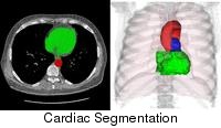 |
Automated Cardiac Analysis Fully automated cardiac segmentation provides foundations for automated measurement of cardiac biomarkers such as the coronary artery calcification content. The goal of this project is to provide fully-automated segmentation and biomarker measurement in the cardiac region in low-dose chest CT. |
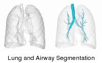 |
Human Airway Analysis Many pulmonary diseases affect the dimensions of airways. By measuring the airway dimensions to a known precision, the clinicians will be able to better diagnose and monitor the patients. |
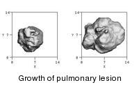 |
Pulmonary nodule analysis on CT scans Lung cancer screening is poised to become a major practice in the near future. As long as patients are undergoing radiation for early lung cancer detection, it is only logical to analyze the same images to diagnose other diseases. Currently, computer algorithms have been developed to measure solid pulmonary nodules, compute growth rates, and determine whether a nodule is malignant or benign from a single scan. |
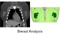 |
Breast Analysis Breast cancer is the most common cancer diagnosed among US women and the second leading cause of cancer death. The purpose of this study was to develop a fully automated framework for breast analysis on low-dose chest CT (LDCT), which consists of breast segmentation, breast density assessment for female subjects, gynecomstia detection for male subjects and breast mass detection. |
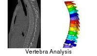 |
Vertebra Analysis Vertebra analysis including individual vertebra segmentation and labeling, as well as closed cortical surface segmentation of the veterbral body, serves as the first step to bone mineral density assessment, osteoporosis detection and compression fracture detection from low-dose chest CT, thereby providing valuable information with respect to the bone health. |
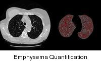 |
Automated Emphysema Quantification CT scan data allows for the measurement of the anatomical basis of emphysema and is being used to determine disease state and progression in clinical cases. Many metrics have been proposed to allow for automated quantification. This research is primarily focused on the analysis of variation of the most commonly used measures to better reflect true progression of disease versus the natural variation present in these measures. |
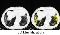 |
Automated Early-Stage ILD Identification Fully-automated algorithms are under development to identify early-stage Interstitial Lung Diseases (ILDs) from chest CT scans using image features and diseased tissue spatial distribution. |
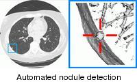 |
Automated nodule detection: Sergei Fotin Examining three-dimensional lung CT images for tiny early-stage nodules is a very tedious and time consuming task for the radiologist. The main goal of this research work is to design and validate an automated system that will detect all cancers without raising too many false alarms. |
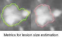 |
Metrics for lesion size estimation: Metrics Group Accurate and reliable measurement of pulmonary nodule size from CT scans has an important role in computer assisted evaluation of lung lesions. This research project aims at studying the reciprocal relationships between different size estimation methods, investingating both the differences in the actual sizes reported and the effects on readers' variability. |
| Neuron 2D-3D Image Registration An automated algorithm is in development to register the in vivo measured individual neurons to those in the histologically cleared brain images. | |
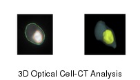 |
3D Optical Cell-CT Analysis Fully-automated algorithms have been developed to segment the nucleus and cytoplasm regions from 3D single cell CT images for cell evaluation such as cell typing. |
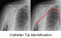 |
Catherter Tip Location Identification: Brad Keller Devolpment of a CAD system to allow for more rapid identification of catheter tips and their location in standard PA chest radiographs. This has applications in intesive care unit reading settings where there is a need to increase throughput due to the both volume of scans and the resultant time-consuming nature of the reads. |
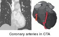 |
Coronary arteries in CTA: Sergei Fotin Decision making in heart disease diagnosis relies heavily on the automated analysis of cardiac images. Here we develop a method for computerized extraction of coronary arteries from CT angiography data. |
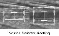 |
Vessel Measurement in Intravital Microscopy: Jaesung Lee Measuring the vessel diameters over time is a crucial step in studying the effect of various stimuli on the width of blood vessel nearby. Automatic measurement provides for fast and accurate tracking of vessel diameter. |
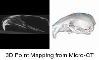 |
3D Point Mapping from Mouse Skull Micro-CT: Jaesung Lee Micro-CT scans allow for the researchers to study phenotypes of small mice. A convenient tool is developed for mapping a 3D point on the mouse skull surface from micro-CT. The mapped points may be used for calculating various dimensions of the skull. |
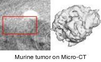 |
Murine lung tumor measurement on Micro-CT scans: Artit Jirapatnakul Mouse models are valuable for studying thedevelopment of human cancer and genetic diseases. Micro-CT scans enableresearchers to monitor the progression of disease starting from a much earlierstage than possible by observing symptoms. Algorithms are being developed toaccurately and robustly measure lung tumors in from micro-CT scans. |
|
GGO Characterization: Andrew Browder, Artit Jirapatnakul Improvements in CT technology and increasing number of accumulated cases have allowed radiologists to find a new type of abnormality, referred to in the literature as GGO or subsolid nodule. These abnormalities have radiological appearances and malignancy rates that are different from those of solid nodules. Characterization of the subsolid nodules could lead to better knowledge about their evolution and better screening protocols | |
|
Dysplastic Hips in Post-Natal Femur: Wendy Vandenberg-Foels Osteoarthritis is the leading cause of adult disability in the United States. Approximately 40% of idiopathic osteoarthritis (OA) cases can be attributed to developmental dysplasia of the hip (DDH). While the endstage OA has been the subject of much research, little is known, about the early postnatal development of the dysplastic hip. The objective of this research is to quantitatively describe the temporal development of normal and dysplastic hips | |