Cardiac Segmentation and Biomarker Measurement
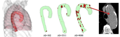
Cardiac health assessment is beneficial for lung cancer screening participants since they are also typically at high risk for cardiovascular diseases. Fully-automated computer algorithms have been developed to segment and measure disease related biomarkers in the cardiac region from low-dose non-contrast chest CT. First, aorta, heart region and the pulmonary trunk region are segmented using rule-based approaches. Then related biomarkers such as the aortic calcification, coronary artery calcification and cardiac visceral fat are segmented and measured. The aorta segmentation algorithm [1] consists of three stages: seed point detection in the aortic lumen at the carina level; cylinder-tracking [2] to establish an aortic centerline; a triangular mesh based model for surface refinement. The algorithm was quantitatively evaluated on 60 CT scans and achieved an average Dice Similarity Coefficient (DSC) of 0.93 compared to manually marked boundaries [1]. The aortic diameter profile is also computed based on the segmented volume. Aortic calcification is extracted from the segmented aorta mask region and measured using an Agatson-weighted score, mass score and volume score. The automatically measured aortic calcification had a high agreement with that extracted from the manual markings (Pearson correlation coefficient R=0.99 using the calcium scores) [3] (see Fig. 1).
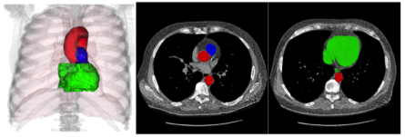
Figure 1. Visualization of segmentation results. Left: segmented aorta (red) with lungs (light pink) and bones (light grey); right: segmented aortic calcification (red) with aorta (light green) in three different cases and their Agatston-weighted scores (AS). The pulmonary trunk region is segmented in the region adjacent to the ascending aorta using an algorithm similar to that used for aorta segmentation [4]. The algorithm was evaluated on 45 manually marked images and achieved an average DSC of 0.88. The heart region is defined by constraints from adjacent organs such as the lungs, aorta and bony structures (see Fig. 2).
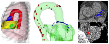
Figure 2. Segmented aorta (red), heart (green) and pulmonary trunk (blue) in coronal view (left image) and two axial slices (middle and right images). Cardiac biomarkers including the cardiac fat content and coronary artery calcification are also segmented [5-6]. Cardiac fat content is associated with cardiovascular diseases. Coronary artery calcification (CAC) content is one of the strongest indicators of coronary heart diseases. Cardiac visceral fat is adjacent to the heart region and constrained by aorta, lungs and sternum. The algorithm was evaluated on 45 manually marked images and achieved an average DSC of 0.93. CAC is segmented based on their relative spatial locations to the segmented aorta, heart, pulmonary trunk and lungs. The algorithm was evaluated on 45 CT scans with manual markings and the correlation between the automated calcium scores and manual reference scores was 0.96 (Pearson correlation coefficient R) (see Fig. 3). The CAC algorithm is further extended to label CAC by their association with the main arteries.
Figure 3. Segmented cardiac visceral fat in yellow (left image), aortic calcification in red and coronary artery calcification in blue (middle and right images).
|