Breast Analysis
Shuang Liu
 Breast cancer is the most common cancer diagnosed among US women, accounting for
nearly 1 in 3 cancers, and the second leading cause of cancer death. Breast
density has been shown to be an independent risk factor of breast cancer, hence
leading to legislation mandating women to be informed of their breast density
reported on mammograms in many states throughout the United States. Gynecomastia
is the most common finding during imaging of the male breast and accounts for most
cases of male breast enlargement, which has been found in more than 57% of men
older than 44 years.
Breast cancer is the most common cancer diagnosed among US women, accounting for
nearly 1 in 3 cancers, and the second leading cause of cancer death. Breast
density has been shown to be an independent risk factor of breast cancer, hence
leading to legislation mandating women to be informed of their breast density
reported on mammograms in many states throughout the United States. Gynecomastia
is the most common finding during imaging of the male breast and accounts for most
cases of male breast enlargement, which has been found in more than 57% of men
older than 44 years.
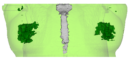
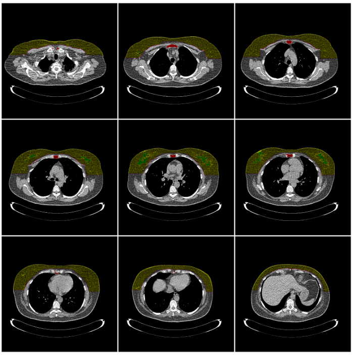
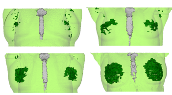
The purpose of this study was to develop a fully automated framework for breast
analysis on low-dose chest CT (LDCT), which consists of the following four
stages:
- The whole breast is segmented, where the fibrogladular tissue and fat tissue
contained in the breast region are also identified.
- For women subjects, the breast density is quantified by classifying the
breast composition into four categories following the Breast Imaging Reporting
and Data System (BI-RADS).
- For man subject, gynecomstia detection is performed based on the volume of
the detected fibrogladular tissue.
- Breast mass is detected base on the analysis of the breast composition.
 The breast region segmentation framework was validated with 1270 LDCT scans,
with 96.1% satisfactory outcomes based on visual inspection [2]. The segmentation
was also quantitively evaluated with 20 LDCT scans by comparing to the ground
truth manually annotated by a radiologist on one axial slice and two sagittal
slices for each scan. The resulting average Dice coefficient is 0.880 with a
standard deviation of 0.058 [1].
The breast region segmentation framework was validated with 1270 LDCT scans,
with 96.1% satisfactory outcomes based on visual inspection [2]. The segmentation
was also quantitively evaluated with 20 LDCT scans by comparing to the ground
truth manually annotated by a radiologist on one axial slice and two sagittal
slices for each scan. The resulting average Dice coefficient is 0.880 with a
standard deviation of 0.058 [1].
 100 scans with satisfactory segmentation were randomly selected for the
validation of breast density measurements, and 91% scans obtained automated
density assessment consistent with subjective reading of an experienced
radiologist [2].
100 scans with satisfactory segmentation were randomly selected for the
validation of breast density measurements, and 91% scans obtained automated
density assessment consistent with subjective reading of an experienced
radiologist [2].
Presentations and Publications
- Liu, S., Salvatore, M., Yankelevitz, D. F., Henschke, C. I., & Reeves, A. P.
Segmentation of the whole breast from low-dose chest CT images
Proceedings of SPIE Medical Imaging,94140I-94140I, March 2015.
- Liu, S., Margolies, L., Xie, Y., Yankelevitz, D. F., Henschke, C. I., &
Reeves, A. P. Fully automated Breast density assessment from low-dose chest CT
Proceedings of SPIE Medical Imaging, submitted, March 2017.
List of Current Research Projects
|
